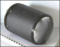
Philips 1964 - 6 Inch Image
Intensifier
|
|
|
|
|
The first image intensifiers in Radiology
were introduced by Westinghouse (the Fluorex
) in 1952, followed by the Philips intensifier, and by the French made “Fluoricon” of General Electric by “ |
|
The above tube is metal protected and was
acquired in 1964. It has a cesium iodide input screen, and boasts an image
intensification factor of about 1000. The bright image on the small output
phosphor could either be viewed through a periscope,
or transmitted through an elaborate optical system to
a TV pick-up tube
to be viewed on a TV monitor. It suffered however, quite often, of a small
central bright spot which had to be eliminated by long gettering.
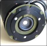
In the picture, above right, this 6”
tube is shown beside a relatively modern 9” Philips intensifier. |
|
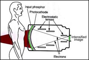
Picture adapted from
“Introduction to Medical Radiographic Imaging”, Eastman Kodak Company, 1993,
p162. |
| Go to Category Index | ||
| Go to Main Page |